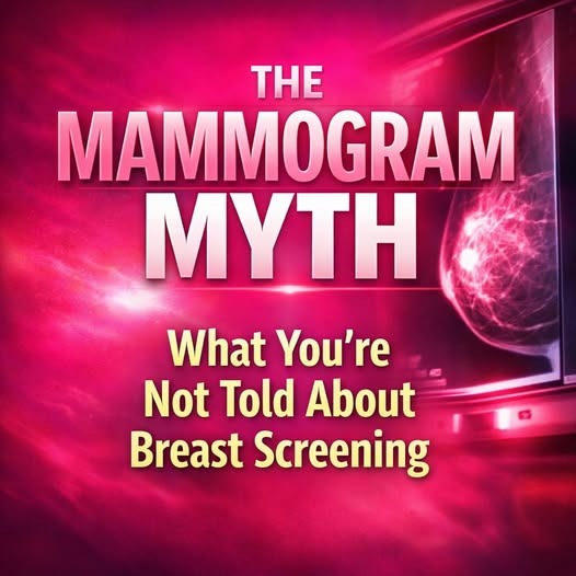
It’s become a routine recommendation — part of mainstream health protocol for women over 40 (and increasingly younger). It’s promoted as the gold standard in catching breast cancer early. And for some, it does detect a mass that leads to timely treatment.
But beneath the surface of this widely accepted test lies a more complex reality — one that deserves full transparency, not fear-driven compliance.
Because when you understand how mammograms really work, their risks, their limitations, and what they can (and can’t) see — you begin to see a bigger picture. One where informed decisions, personalized care, and terrain awareness matter more than one-size-fits-all screening.
Let’s look deeper:
A mammogram is a low-dose X-ray image of the breast that attempts to identify abnormal masses, calcifications, or structural changes that could indicate cancer.
During the procedure:
• The breast is compressed tightly between two plates
• X-ray radiation passes through tissue
• Denser areas (like tumors or calcifications) appear as white spots
It’s a mechanical, structural test — and while it may detect a suspicious lump, it cannot diagnose cancer on its own. It can only identify an abnormality. Further testing (usually biopsy, ultrasound, or MRI) is required to confirm malignancy.
It’s also important to understand that a suspicious area on a mammogram does not always equal danger. And a clear mammogram doesn’t always mean safety.

Check the first comment😲👇

The mysterious metallic object that intrigued the Internet: do you know what it was used for?

Amazon Raises Prices for Ad-Free Streaming Tier, Rebrands It Prime Video Ultra

A Resident Evil Requiem fan who registered a secret in-game website says he will return the site's server to Capcom for free. Provided that the company contacts him

"This Little Dude Spied On Me For 10mins" Nosy little guy, ain't he?